Issue 52 Summer 2021
Issue 52 Summer 2021
Bringing The Latest In Paediatric Endocrinology To You
2344 Likes
Editorial
Welcome
Thanks to our excellent speakers, we are fortunate in this issue of ESPE News to have
insights into several of the talks you will be able to enjoy at ESPE 2021 Online. On page 6, Ali Abbara and Waljit Dhillo review our
understanding of kisspeptin’s role in puberty, with exciting potential developments in its therapeutic and diagnostic use. Meanwhile, on page 7, Peter Kühnen examines the melanocortin
4 receptor (MC4R) agonist setmelanotide as a treatment option in rare obesity syndromes. He explains the capacity of MC4R agonists to activate
different downstream signalling cascades (‘biased signalling’) and therefore elicit a range of effects. Supporting transgender/gender diverse youth
remains a complex and topical area of healthcare. Stephen Rosenthal discusses the associated issues, which he will address in his forthcoming presentation (page 8).
You can find out more about ESPE 2021
Online at www.eurospe.org/espe2021online.
As always, your contributions will form a central part of the meeting, so please make sure to submit your abstracts by 10 May 2021.
On page 4, we are extremely pleased to have contributions from colleagues in India about their lives in the time of COVID. Researchers
Anuradha Khadilkar and Vandana Jain reflect on the pandemic’s impact on their work with patients and other aspects of their research and daily
lives.The rest of the issue is bursting with the opportunities and support available to you from
ESPE. These extend from grants and committee vacancies to the prospect of future events such as ESPE Schools and the postponed ESPE Science
Symposium. Read on to learn more!We thank all this issue’s contributors for writing for us at such a busy and stressful time.
We wish them, you, and all your families and friends, health and peace in the coming months.
Sarah Ehtisham
Editor, ESPE News
[email protected]
News
Intro
Follow ESPE
online…
Keep an eye on
the latest ESPE news
and activities at
www.eurospe.org
You can also follow
ESPE on Facebook
and Twitter/EuroSPE
News
ESPE 2021 Online
Register today and benefit from the early bird registration fees for ESPE 2021 Online. www.eurospe.org/espe2021online
News
Marie-Jose Walenkamp remembered
We are sad to report the untimely death of Marie-José Walenkamp (The Netherlands), an active ESPE member, excellent clinician and scientist with paradigm-changing research contributions, who worked primarily on genetic causes of disturbed growth. Marie-José also taught paediatric endocrinology, nationally and internationally. Our thoughts and sympathy are with her family, friends and colleagues at this very difficult time. You will find a full obituary at www.eurospe.org/news/ article/16117/obituary-marie-josé-walenkamp
News
Early Career Scientific
Development Grant
This ESPE grant supports personal development though a short visit to an external laboratory/hospital/institute. While international travel restrictions are in place during the pandemic, it can be used for a research period of up to 3 months at your home location. It may be used to finance the visit of an outside expert to your institution, to provide essential guidance, consultation or advice. The grant is intended to help you: • learn a new technique • troubleshoot existing methods • discuss new methods/ techniques in a research seminar • compile statistics on joint research projects • test samples in the framework of a joint research project. During my stay I learned PCR, site-directed mutagenesis, transfection, luciferase assay and Western blot assay. All the techniques were taught to me one-on-one, and I performed them under supervision. I also had a chance to present my study in a team meeting, and attended seminars at the research centre.” Sare Betul Kaygusuz (Turkey), former grant recipient Apply by 30 September 2021
News
Clinical Fellowship
2021 is the 30th anniversary of the ESPE Clinical Fellowship programme. Since 1991, it has supported more than 250 trainees from over 30 countries across four continents. The ESPE Clinical Fellowship Spring Initiative in April provided an online forum for former and current ESPE Clinical Fellows, hosts and tutors, as well as the applicants for 2020 who had not yet been through the selection process.
The diverse 3.5-hour programme attracted over 100 fellows and featured state-of-the-art presentations, tutors’ patient presentations and fellows’ presentations (cases and research). It was well-received, as illustrated by the feedback we received: Wonderful cases and great presentations by the fellows and teachers, with lots of learning points. Thank you ESPE for this initiative and its beautiful execution.” Thanks for arranging such an excellent learning platform during this pandemic situation. I also thank all presenters for presenting interesting cases.” All ESPE members can view the recorded content at www.eurospe.org/members/clinical-fellowship-spring-initiative Winter School 2021 Online 27 February–2 March 2021 Cancellation of the 2020 Winter School at very short notice, due to the pandemic, strengthened ESPE’s resolve to ensure it took place in 2021, even if not in person. Restructuring by the teaching faculty optimised online interaction. Rehearsals familiarised students and teachers with the web platform.
Approximately 5 hours of continuous teaching was delivered daily for 4 days. Small group teaching sessions used online breakout rooms, lectures were punctuated by polling questions, and audience participation was vigorously encouraged! This was the first ESPE school to be moved online. Feedback certainly suggests it was successful. It has since been followed by the Summer and Diabetes, Obesity and Metabolism Schools in May. While all agreed that an online event was far better than no event, face-to-face education will return as soon as possible, and we hope Winter School will be in Moldova in 2022. Please also look out for adverts for two teaching faculty members shortly.
ESPE members with a passion for training and supporting the next generation of trainees should apply. See the full report at www.eurospe.org/news/article/16126/report-from-espe-winter-school-online-2021 Remaining Schools for 2021 Due to the ongoing pandemic and closure of borders, the ESPE Caucasus & Central Asia School, Maghreb School and ASPED-ESPE Endocrine Academy will be postponed until early 2022, when they can hopefully be held in person. Applicants will be informed accordingly. Find out more at www.eurospe.org/education
News
ESPE e-Learning
The category Calcium and Bone under General Content offers the following five chapters, plus 13 problem-solving cases: • Calcium and bone metabolism • Bone dysplasia • Growth plate maturation • Hypophosphatasia, and • Metabolic bone disease of prematurity. In addition, Bone Disorders within Resource Limited Countries includes a chapter Calcium and Bone Health and two problem-solving cases, all in five languages (English, French, Spanish, Chinese and Swahili). News in e-learning A new chapter, ‘Hypothyroxinaemia of Prematurity’, has been added to the category Thyroid Disorders under General Content. See www.espe-elearning.org Registration is free of charge
Hot topics
Intro
Bringing you recent highlights from the world of research
Hot topics
Hydrocortisone granules for adrenal insufficiency in children
Neumann et al. conducted a prospective study to evaluate the efficacy and safety of hydrocortisone granules in children with adrenal insufficiency and congenital adrenal hyperplasia (CAH). Children with CAH (n=17) or hypopituitarism (n=1) under 6 years of age were followed prospectively for 2 years. 17-Hydroxyprogesterone saliva profiles were used every 3 months to adjust therapy in cases of CAH. Hydrocortisone granules were administered three times a day. The median duration of treatment was 795 days (range 1–872 days). There were 150 follow-up visits. The median (range) daily dose of hydrocortisone (mg/m2 ) at the start of the study was 11.9 (7.2–15.5) (age 2–8 years), 9.9 (8.6–12.2) (age 1 month–2 years) and 12.0 (11.1–29.5) (age <1 month). At the end, values were 10.2 (7.0–14.4), 9.8 (8.9–13.1) and 8.6 (8.2–13.7) respectively. Growth remained normal and no adrenal crises were observed. Thus, accurate dosing appears safe and without increased risk of adrenal crisis. Read the full article at Neumann et al. 2021 Journal of Clinical Endocrinology & Metabolism 106 e1433–e1440
Hot topics
CXM: a real-time marker of height velocity
Coghlan et al. studied the utility of a bone growth by-product, collagen X biomarker (CXM), as an indicator of height velocity (HV). They have suggested the working reference ranges and charts for boys and girls. Blood samples from 302 healthy children (163 boys) and 10 healthy adults were analysed. Of the 961 CXM measurements, 432 were plotted by age, and sex-specific reference ranges were determined. The serial values of CXM from 116 participants were plotted against observed HV. The plots of CXM in ng/ml measured over the ages matched roughly with the standard HV plots. There was a strong correlation (r=0.80, P<0.001) between blood CXM and conventional HV. The CXM levels showed the pubertal growth spurt in both girls and boys. There was no difference in CXM levels in matched serum, plasma and dried blood spot samples. However, the non-availability of a standard CXM assay, its diurnal variation and poor reliability of single point assessment are potential limitations in the widespread clinical use of CXM as a marker for HV
Hot topics
Congenital hypothyroidism: consensus guidelines
This update to the 2014 guidelines is written by the Endo-ERN (the European Reference Network on Rare Endocrine Conditions) and endorsed by ESPE and the European Society of Endocrinology. The guidelines emphasise screening for congenital hypothyroidism (CH) using thyrotrophin (TSH) for early detection to prevent neurodevelopmental delay. Free thyroxine (T4) measurement can be added to screen for central CH, where feasible. There are suggested thresholds for treatment (low free T4 or confirmatory TSH >20mU/l) or interval re-evaluation (TSH 6–20mU/l). Levothyroxine treatment should be started at no later than 2 weeks of age, at 15µg/kg per day, with lower doses for mild CH. The initial goal is a rapid increase in circulating thyroid hormone and normalisation of TSH. Once treatment is established, a thyroid function test is recommended before or 4 hours after a feed. The dose should not be reduced for a raised free T4 level without TSH suppression or clinical signs of overtreatment. When doses are adjusted, re-evaluation in 4–6 weeks is recommended. If definitive diagnosis of permanent CH has been made, then re-evaluation of the hypothalamic–pituitary–thyroid axis after 2–3 years of age is indicated, particularly in children with a gland in situ, and in those with presumed isolated central CH. The guidelines also cover outcomes, prognosis and genetics
Hot topics
Neutropenia in hyperthyroidism
Neutropenia may be a sign of new-onset hyperthyroidism. Finding neutropenia in a newly diagnosed hyperthyroid patient can represent a therapeutic dilemma, as severe neutropenia or agranulocytosis (absolute neutrophil count (ANC) <0.5×10⁹ /l) is a well known adverse effect of antithyroid drugs (ATDs). This systematic review and meta-analysis by Scappaticcio et al. aimed to assess the prevalence, degree and response to treatment of neutropenia in hyperthyroid patients. In total, 13 studies involving 1144 patients (991 with Graves’ disease), aged 37–46 years, were included. Mild to moderate neutropenia (ANC 0.7–1.75×10⁹ /l) occurred in 103 newly diagnosed, untreated, patients with Graves’ disease. In all of them, hyperthyroidism treatment (ATDs, prednisone or radioiodine) led to resolution of neutropenia. No infectious diseases or events of ATD-induced agranulocytosis were recorded amongst patients with neutropenia. The authors conclude that Graves’ disease is associated with neutropenia in about 10% of cases. Mild neutropenia is not a contraindication for ATD treatment, as hyperthyroidism treatment can lead to neutropenia resolution. Read the full article at van Scappaticcio et al. 2021 Clinical Endocrinology 94 473–483
Feature
Feature
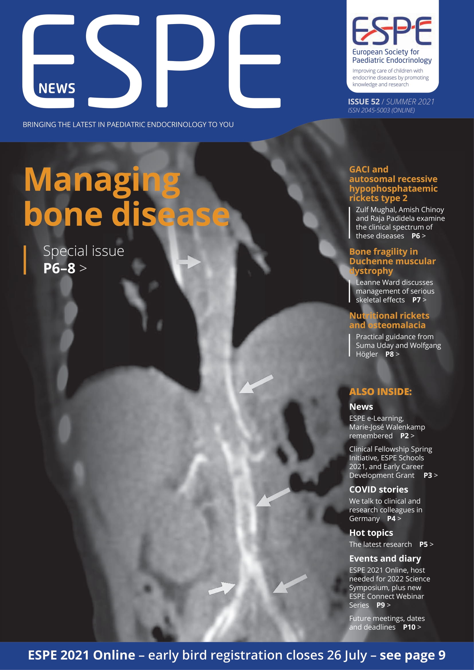
GACI and autosomal recessive hypophosphataemic rickets type 2
Generalised arterial calcification of infancy (GACI) is a devastating condition associated with significant mortality and morbidity.1,2 It is characterised by stenosis of large and medium-sized arteries arising from calcification and intimal proliferation. Antenatal ultrasound scans may show hyperechogenic signals arising from calcification of heart valves and blood vessels, polyhydramnios, pericardial effusion, cardiac ventricular hypertrophy and dysfunction. Affected infants often die in utero. Approximately 50% of infants who are born alive die in the first 6 months of life from myocardial infarction, congestive heart failure, hypertension, strokes and ischaemic organ damage, arising from vascular stenosis. Aetiology GACI type I arises from homozygous loss of function mutations in the ENPP1 gene, which result in deficiency of ectonucleotide pyrophosphatase phosphodiesterase 1 (ENPP1), an extracellular transmembrane enzyme. ENPP1 deficiency leads to failure of hydrolysis of extracellular ATP into inorganic pyrophosphate (PPi) and AMP. PPi is a physiological inhibitor of calcification, while AMP and its metabolite adenosine contribute to arterial intimal hyperplasia.3 GACI type II is an autosomal recessive disorder arising from loss of function mutations in the ABCC6 (ATP-binding cassette, subfamily C, member 6) gene, which more typically causes pseudoxanthoma elasticum (PXE).4 Patients with PXE, who also have low plasma PPi levels, develop progressive calcification of elastic fibres in the skin, eyes and arterial walls. Clinical features of PXE include skin laxity, yellowish skin papules around the neck and flexural regions, retinal angioid streaks and arterial calcification causing myocardial infarction and strokes. Clinical features of PXE have also been reported in survivors of GACI type I, suggesting that there is clinical overlap between the two subtypes of GACI. Skeletal effects Calcification of joints and deafness occur in patients with GACI types I and II, while the majority of survivors of GACI type I go on to develop autosomal recessive hypophosphataemic rickets type 2 (ARHR2). ARHR2 secondary to ENPP1 deficiency is mediated by elevated plasma fibroblast growth factor-23 (FGF23) levels, leading to renal phosphate excretion and impaired intestinal phosphate absorption secondary to low serum 1,25-dihydroxyvitamin D (1,25(OH)2 D) levels
Patients with ARHR2 present with clinical features of rickets, including short stature and lower limb deformities, e.g. genu valgum or genu varum. The child may walk with a waddling gait, due to proximal myopathy and coxa vara that often accompanies genu varum. An older child may complain of easy fatigability and bone pain. Patients with ARHR2 have an inappropriately low maximal reabsorption rate of phosphate per unit volume of glomerular filtrate for their prevailing, low age-appropriate serum phosphate value, a normal serum calcium value and a raised serum alkaline phosphatase level for their age. Serum parathyroid hormone level is usually normal or mildly elevated due to inappropriately low serum 1,25(OH)2 D levels.5,6 The serum level of 25-hydroxyvitamin D is usually normal and intact plasma FGF23 level is elevated. A radiograph of a wrist or a knee will show classical radiological changes of rickets: widening, cupping and fraying of metaphyses. ARHR2 due to homozygous loss of function mutations in the ENPP1 gene also arises in patients without features of GACI, such as arterial stenosis secondary to calcification and intimal hyperplasia. Treatment In the absence of a cure, treatment of GACI is essentially supportive. Bisphosphonates, especially earlier generation non-nitrogen-containing bisphosphonates such as etidronate, are non-hydrolysable PPi analogues, which have been used as off-label medications to reduce arterial calcification. There is no convincing evidence that bisphosphonates influence morbidity and mortality in infants with GACI. Furthermore, bisphosphonates do not prevent stenosis of arteries resulting from intimal proliferation. Rickets due to ARHR2 is treated by administration of phosphate salts four or five times a day, along with active analogues of vitamin D: calcitriol (1,25(OH)2 D) or alfacalcidol (1-hydroxycholecalciferol).6 In preclinical studies, ENPP1 enzyme replacement therapy using the recombinant ENPP1 fusion protein successfully prevents the development of calcification and intimal proliferation.3,7 This treatment improved survival in animal models of GACI type 1.7,8 It also prevented the development of ARHR2.9 Trials of ENPP1-Fc replacement therapy in humans with ARHR2 and GACI will be starting shortly. Zulf Mughal, Amish Chinoy and Raja Padidela Department of Paediatric Endocrinology, Royal Manchester Children’s Hospital, and Faculty of Biology, Medicine and Health, University of Manchester, UK Conflict of interest ZM has received consultancy fees from Inozyme Pharma, Inc. He is also a medical advisor to GACI Global.
Feature
Bone fragility in Duchenne muscular dystrophy
Patients with Duchenne muscular dystrophy suffer serious skeletal consequences. Leanne Ward discusses management strategies and unmet needs
Duchenne muscular dystrophy (DMD) is an X-linked recessive disorder characterised by a progressive myopathy arising from loss of function mutations in the dystrophin gene. Without treatment, ambulation is lost by 10 years of age on average, and death due to cardiorespiratory failure occurs about a decade later, unless ventilated. High dose glucocorticoid (GC) therapy is the only approved treatment for all patients with DMD, prolonging ambulation on average by 2 years, with modest extension of life expectancy. However, significant side effects limit GC use. These include linear growth failure, delayed puberty, obesity and fragility fractures. The search for GC-sparing therapies to arrest or significantly attenuate the declining muscle function is one of the most intense areas of study in paediatric medicine the world over. Bone health in DMD Bone fragility in DMD is a potentially severe and disabling consequence of the myopathy and its treatment. The skeletal phenotype is driven by disease-related perturbations in the muscle–bone unit (including lack of periosteal apposition, due to mechanical under-stimulation, and abnormal muscle–bone ‘cross-talk’), as well as the osteotoxic effects of GCs. Disease-related issues drive the long bone fractures, which are exacerbated by GC therapy, whereas vertebral fractures are more common with GCs. The symptomatic vertebral fracture prevalence in GC-treated patients is >50%, with vertebral fractures occurring in almost a third of patients in the 4 years following GC initiation. Since vertebral fractures are typically asymptomatic in their early stages, studies to date have probably underestimated the total vertebral fracture burden in DMD.
The consequences of osteoporosis can be devastating to patients and families, including premature, permanent loss of ambulation, and death due to fat embolism syndrome. Long bone fractures have been reported prior to GC therapy, and even prior to diagnosis, while vertebral fractures typically first appear about 1–2 years (but as early as 6 months) following GC initiation. When vertebral fractures are undetected or untreated, boys are at risk of the ‘vertebral fracture cascade’, involving more painful, numerous and severe collapse that occurs after an initial vertebral fracture event. Prevention of this phenomenon is one of the primary goals of treatment, particularly since potential for ‘medication-unassisted vertebral body reshaping’ is limited in DMD. Disease management The Centres for Disease Control DMD Task Force published Care Considerations in the Lancet Neurology in 2018. These advise that periodic spine surveillance
(by X-ray or dual-energy X-ray absorptiometry) should be initiated around the time of diagnosis or GC prescription, using a validated vertebral fracture quantification method. The 2018 DMD Care Considerations also recommend starting intravenous bisphosphonate therapy after a single long bone or vertebral fracture. With this ‘secondary prevention approach’, back pain and further vertebral collapse are mitigated to a greater extent following early, rather than late, intervention. Despite early intervention with intravenous bisphosphonate therapy, long bone fractures may still occur, and vertebral fractures may still progress (particularly when in more advanced stages of collapse at bisphosphonate initiation). This raises the question of whether the aggressive osteoporosis can be halted if intravenous bisphosphonate therapy is given before first ever fractures. This question remains unanswered to date. Intravenous bisphosphonates have the greatest benefit at trabecular-rich sites such as the spine, have less of an effect on compact bone (i.e. long bone diaphyses), and do not augment periosteal circumference. Such observations point to the need for more effective strategies, which can be approached theoretically from different angles, including disease-modifying therapies that are less toxic to bone than GCs, dual action (combined anti-resorptive/anabolic) therapy, or sequential (anabolic followed by anti-resorptive) treatment. These strategies are as yet untested, and provide a blueprint for future study designs to effectively prevent osteoporosis and its consequences in this context. Leanne M Ward Professor of Pediatrics, Tier 1 Research Chair in Pediatric Bone Diseases, University of Ottawa, and Pediatric Endocrinologist, Children’s Hospital of Eastern Ontario, Canada
Feature
Nutritional rickets and osteomalacia
Suma Uday and Wolfgang Högler provide a practical guide to managing these widespread yet preventable conditions.
Rickets and osteomalacia result from defective mineralisation of the growth plate and the preformed osteoid respectively. Solar vitamin D and/or dietary calcium deficiency are the leading causes of rickets and osteomalacia, constituting a major global health problem. Clinical presentation and risk groups The pathophysiology is complex.1 The clinical presentation of rickets/osteomalacia can be associated with deficiencies in phosphate and/or calcium. Presentation depends on age, with symptomatic hypocalcaemia (seizures, tetany) predominating in the rapid growth phases of infancy and adolescence.2 The most serious but rare consequence of hypocalcaemia is heart failure due to dilated cardiomyopathy in infants.3 Children with severe or prolonged deficiency can present with poor growth, stunting and bowing deformities.2 However, many cases of osteomalacia may go undiagnosed due to non-specific symptoms such as fatigue, malaise and muscle or bone pain.4 The population most at risk includes pregnant women, infants, individuals with dark skin residing at high latitudes with low ultraviolet B availability, sun-avoidance behaviour and malabsorption or restricted dairy diet.5 Definitions of deficiency Vitamin D. Serum 25-hydroxyvitamin D (25OHD) concentrations <30nmol/l (12µg/l) represent deficiency, while >50nmol/l (20µg/l) represents sufficiency, and 30–50nmol/l (12–20µg/l) is insufficiency.5 Dietary calcium. Dietary calcium intake >500mg/day is classed as sufficient to prevent rickets. An intake <300mg/day is classed as deficient in all individuals except infants. For infants aged 0–6 months, 200mg/day is classed as sufficient; for those aged 6–12 months, 260mg/day is sufficient.5 Investigations and diagnosis Rickets is a radiological diagnosis, comprising cupping and fraying of the metaphyses with widening of the growth plates and radiological osteopenia. Osteomalacia may manifest with Looser’s zone fractures on radiographs. However, definitive diagnosis requires histomorphometric confirmation.4 Nonetheless, rickets/osteomalacia can be suspected based on the predisposing risk factors, dietary history and clinical presentation, and the diagnosis established on the typical biochemical markers: high serum parathyroid
hormone (PTH) and alkaline phosphatase (ALP); low phosphate.4,6 Elevation of PTH is an early sign of vitamin D and/or calcium deficiency.6 A drop in serum calcium only occurs at a very late stage and is a poor indicator of body calcium stores.6 On the contrary, serum 25OHD levels are stable and a good indicator of vitamin D status. Investigations in infants presenting with symptomatic deficiency must include chest radiographs, electrocardiogram and echocardiogram to assess cardiac status.3 Treatment and prevention It is essential to differentiate between simple deficiency states (requiring supplements) and full-blown rickets/ osteomalacia (requiring treatment). Pharmacological management of rickets incorporates treatment doses of vitamin D with or without calcium. Both ergocalciferol (D2) and cholecalciferol (D3) can be used for daily oral supplementation. However, D3 is preferred for stoss therapy (i.e. a single high dose treatment), due to its longer half-life.5 Treatment doses of D2 or D3 are administered for a minimum of 12 weeks, and calcium supplementation until adequate dietary calcium intake can be established. For age-based doses of vitamin D for daily treatment and stoss therapy see the Table.5 Stoss therapy should only be used in infants >3 months of age and in situations of non-compliance.5 Any treatment should be followed by lifelong supplementation with maintenance doses unless the underlying risk factors can be mitigated, which is rare. It is imperative to ensure an adequate calcium intake of at least 500mg/day in all ages except infants (see above) through diet or supplements.5 Children presenting with acute or life-threatening hypocalcaemic symptoms warrant intravenous calcium gluconate administration for immediate correction of serum calcium.3 Whilst activated forms of vitamin D (alfacalcidol or calcitriol) can be used in the acute setting to enhance calcium absorption, they do not have a role in the ongoing management of nutritional rickets/osteomalacia. Screening of family members who share the same risk factors is vital.3 From a public health perspective, the focus should be on prevention. Long term strategies should include food-based solutions such as mandatory food fortification7 with vitamin D and/or calcium or biofortification. Additionally, robust supplementation of high risk groups7,8 through monitored national programmes8 is indispensable both in the short and long term, especially in high latitude countries. In conclusion Symptomatic nutritional rickets/osteomalacia, which are preventable, only represent the tip of the iceberg of widespread deficiency. Food fortification and targeted supplementation of high risk groups should be the way forward in tackling this global health issue. Suma Uday and Wolfgang Högler Institute of Metabolism and Systems Research, University of Birmingham, UK, and Johannes Kepler University, Linz, Austria
Events
Lifelong endocrine care through collaboration, discovery and innovation’
Events
ESPE 2021 Online
22–26 September 2021 ‘Lifelong endocrine care through collaboration, discovery and innovation’ Join us virtually this September for the 2021 Annual ESPE Meeting. The ESPE Programme Organising Committee has created an exciting and robust programme, covering basic science, translational research and clinical care, offering you the very best update in the field of paediatric endocrinology. ESPE 2021 Online will deliver 5 days of sessions to support your continued education. Immerse yourself in the latest developments in paediatric endocrinology, whilst networking virtually and collaborating with peers from around the globe. Unlock a world of content with a wealth of sessions including: • 8 Plenary lectures from experts in their fields • 22 Symposium sessions to keep you up to date on developments • 8 Meet the Expert sessions to learn from esteemed endocrinologists • 2 Controversy sessions, triggering discussions between you and our speakers • 2 How Do I….? sessions, on best management of clinical problems.
Events
Could you host the 2022 ESPE Science Symposium?
Would you like to plan and deliver a 1.5-day symposium on an area of paediatric endocrinology? If you or your collaborative group would like to: • showcase the latest scientific knowledge • host topical discussions and • provide networking opportunities for scientists and clinicians then we would welcome your application as a potential local organising committee for the 2022 ESPE Science Symposium. Your meeting topic should be attractive to researchers in basic or translational research, as well as ESPE members developing experimental and clinical research within and beyond paediatric endocrinology. We look forward to receiving your application! Apply by 30 June 2021 Find guidelines and an application form at www.eurospe.org/education/espe-science-symposium
[contact-form-7 id="520" title="Comments"]